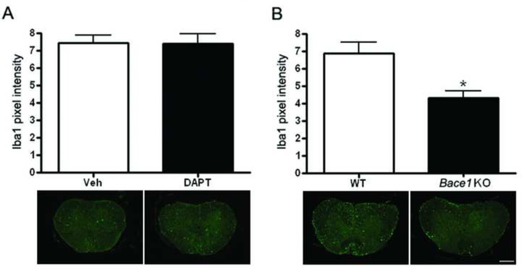
Figure 7. Bace1 KO mice show less microglia after SCI.
Sections form vehicle-treated (Veh, n=6), DAPT-treated (DAPT, n=6), Wild type (WT, n=3), and Bace1 KO (n=3) mice 28 days after injury were stained with Ibal, and staining quantitation was performed using image J. A. No differences in microglial number are observed in Veh and DAPT mice as shown in the graph and the representative images. B. A significant difference (p value = 0.002) in microglial number is observed between the Bace1 KO and the WT mice as shown in the graph and the representative images (Mag. Bar = 500 µm).

