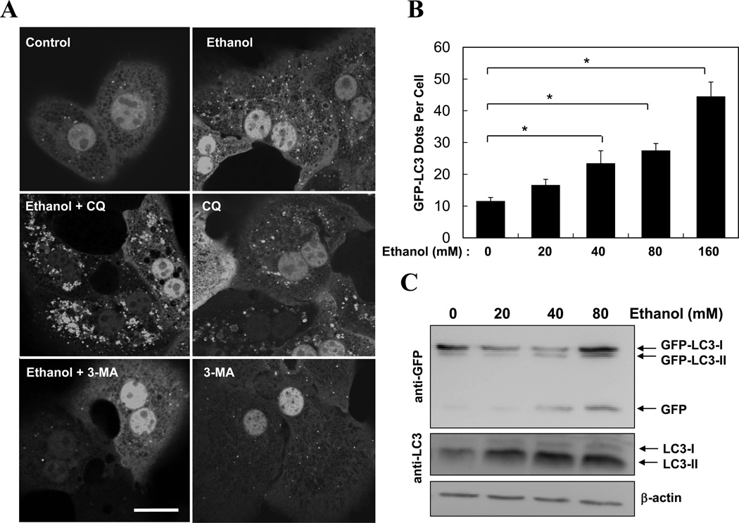
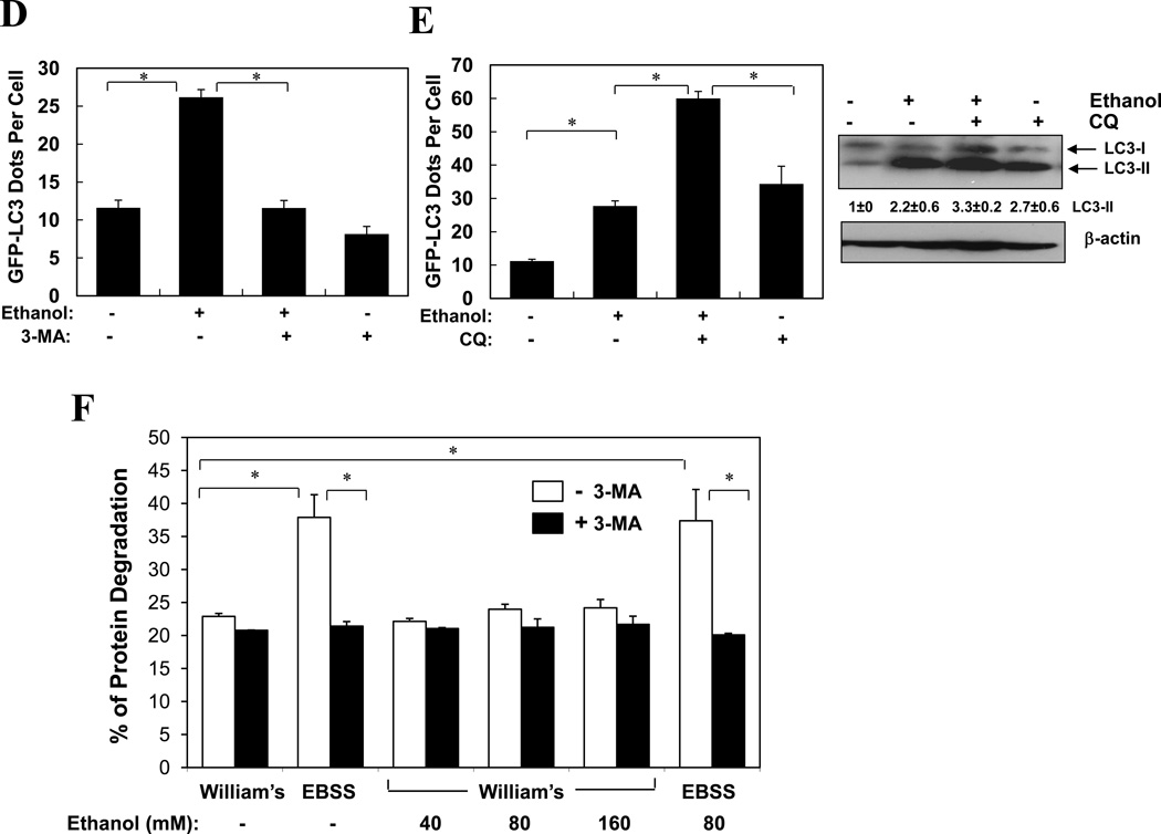
Figure 2. Ethanol induces autophagy in hepatocytes in vitro.
(A–C). Ad-GFP-LC3 infected hepatocytes were treated as indicated (80 mM of ethanol in A) for 6 hours and examined by confocal microscopy (A) or immunoblot assay (C). Scale bar: 20 µm. GFP-LC3 dots (mean+SEM) were quantified for each experiment (n=3). (D–E). Ad-GFP-LC3 infected hepatocytes were treated with ethanol (80 mM, D, 40 mM, E–F) with or without 3-MA (D) or CQ (E, F) for 6 hours. GFP-LC3 dots (mean+SEM) were quantified for each experiment (n=3). Total lysates were subjected to immunoblot assay and densitometry analysis (mean+SEM) of the LC3-II was performed (n=3). (F). Hepatocytes were cultured in William’s medium/10% serum or in EBSS without or with ethanol for 15 hr and long-lived protein degradation was determined (mean+SEM, n=2–3). *: p<0.001.


