
Figure 7. Model of in vitro coordination of bone and vascular tissue development.
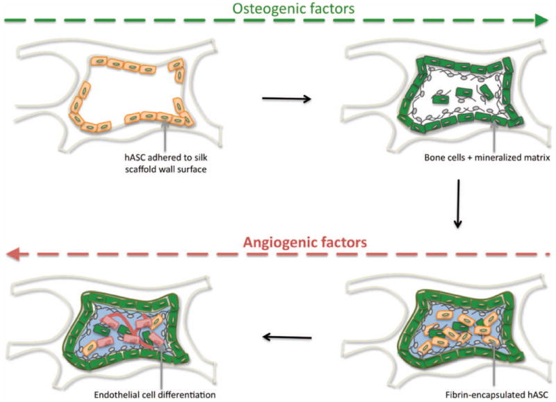
hASC (beige spindle-shape) adhered to silk scaffold wall, sense the hard surface and osteogenic factors provided, stimulate osteogenic differentiation (green cells) and deposition of mineralized matrix (grey). At a second stage, hASC encapsulated in fibrin hydrogel (light blue) are triggered by smooth vasculogenic surface growth factors provided, to undergo endothelial differentiation (red cells).