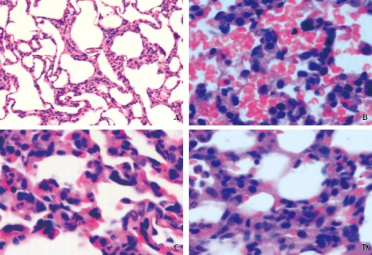
Figure 1.

The histopathological changes of lung by a light microscope. A: the control group (original magnification × 400): normal alveolar spaces, alveolar septum. B: the LPS group (original magnification × 400): the diffuse alveolar septa widened with a lot of red blood cells and inflammatory cell infiltration. C: the LPS + VIP group (original magnification × 400): the focal widened alveolar septum and cavity with a small amount of red blood cells, inflammatory cell infiltration. D: the LPS + VIP + GC group (original magnification × 400): light alveolar hemorrhage, a small amount of inflammatory cells in the alveolar wall.
