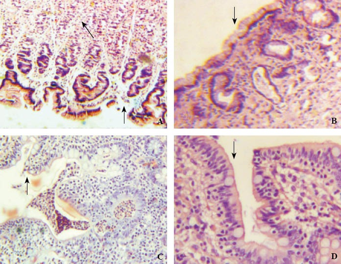
Figure 3.

The pathological changes of gastrointestinal tissue structures observed by a light microscope in the control and hypothermia groups.
A: Gastric mucosa (×100) showing defect of mucosal epithelial cells (black arrow), the arrangement of cells in lamina propria layer loose and disordered, representing ischemic changes (white arrow head) in the control group; B: Gastric mucosa (×100) showing mucosal epithelium complete (black arrow head) in the hypertransfusion group; C: Intestinal mucosa (×100) showing the structure of intestinal villi basically complete, but the arrangement of cells irregular (black arrow) and a plenty of inflammatory cells in the mucosal layer (white arrow) in the control group; D: Intestinal mucosa (×400) showing the structure of intestinal villi complete and the arrangement of cells regular and dense (black arrow) in the hypertransfusion group.
