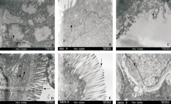
Figure 4.

The pathological ultra-structural changes of gastric tissues observed with an electron microscope in the control and hypothermia groups.
A: Gastric mitochondria were highly swollen; vacuole–like changes with ridges fragmental could be observed (black arrow) in the control group; B: The structure of gastric mitochondria was complete with their ridges relatively clear (black arrow) in the hypertransfusion group; C: Defect of part of intestinal microvilli (black arrow) in the control group; D: In the control group, the arrangement of intestinal microvilli loose (black arrow) and the structure of intestinal mitochondria not distinct with their ridges hazy (black arrow); E: The arrangement of intestinal microvilli dense and regular (black arrow) in the hypertransfusion group; F: The intestinal mitochondria with their ridges manifesting basically clear (black arrow) in the hypertransfusion group.
