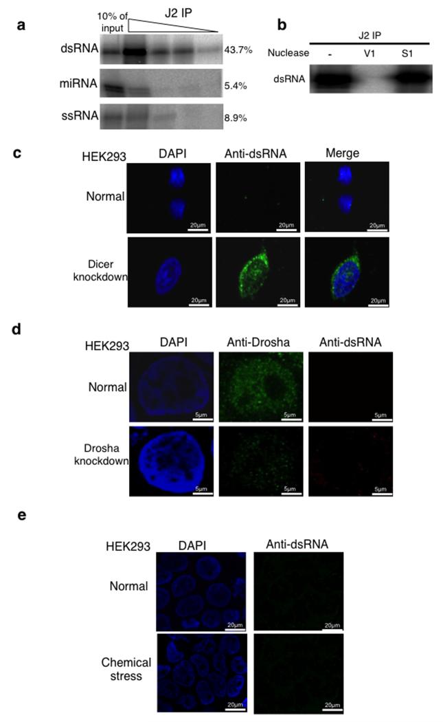
Figure 4. Loss of Dicer leads to accumulation of long dsRNA.
a) Immunoprecipitation analysis of uniformly 32P-radiolabelled dsRNA, miRNA and, ssRNA (See Online Methods) at 10-fold decreasing dilutions using J2 antibody. Signals were visualized and quantitated by PhosphoImager. The level of signal for the first concentration is shown on the right-hand side of the blot and it is expressed as a % of input. This experiment was replicated twice. See Supplementary Fig. 9 for uncropped blot images.
b) IP of dsRNA as in (a) and treated with V1, S1 or not treated. See Supplementary Fig. 9 for uncropped blot images.
c) Immunofluorescence (IF) analysis using J2 antibody showing dsRNA (green) in normal and Dicer knockdown HEK293 cells. Nuclei were stained with DAPI (blue).
d) IF analysis using anti-Drosha and J2 antibodies (green) in normal and Drosha knockdown HEK293 cells. Nuclei were stained with DAPI (blue).
e) IF analysis using J2 antibody on normal cells and cells treated with 2mg/ml acivicin for 48 hours. All experiments described in Fig. 4c-e, were independently replicated three times.

