
Figure 1.
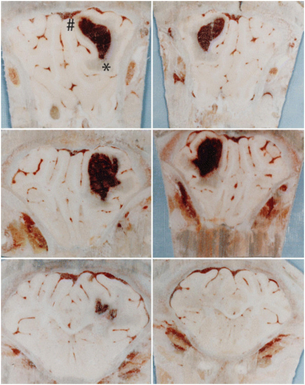
Photomicrographs showing both sides of serial coronal sections (thickness approximately 5 mm) containing hematoma (#) and edematous white matter (*) 24 hours after blood infusion in a pig brain frozen in situ. Perihematomal edema is present as blue-staining translucent regions in white matter adjacent to the hematoma. Evans blue staining is observed throughout the ipsilateral white matter and is indicative of increased BBB permeability and vasogenic edema development. Figure/legend reprinted with permission from Journal of Neurosurgery[11].