
Figure 3.
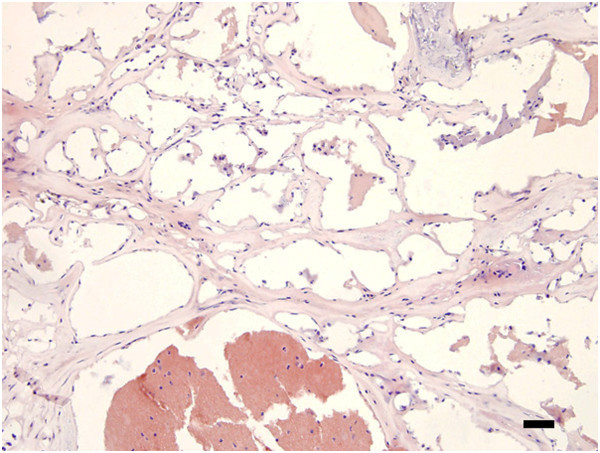
Hematoxylin & eosin staining of a section from the brain of a patient with cerebral cavernous malformation (type CCM3). Note the multiple blood vessels with little or no intervening parenchymal tissue. Scale bar = 200 μm.
Official websites use .gov
A
.gov website belongs to an official
government organization in the United States.
Secure .gov websites use HTTPS
A lock (
) or https:// means you've safely
connected to the .gov website. Share sensitive
information only on official, secure websites.
Hematoxylin & eosin staining of a section from the brain of a patient with cerebral cavernous malformation (type CCM3). Note the multiple blood vessels with little or no intervening parenchymal tissue. Scale bar = 200 μm.