
Figure 25.
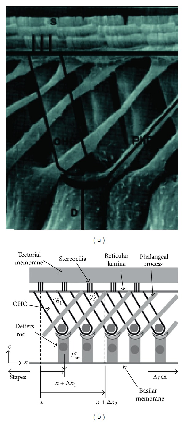
(a) Anatomical scan of a longitudinal view of the organ of Corti of a mole rat cochlea with a representative OHC, Deiters' rod (D), phalangeal process (PhP), and stereocilia bundle (S). (b) Schematic of the longitudinal view of the organ of Corti, showing the tilt of the OHCs based on (a) [160] (reprinted from Biophysical Journal, 100, Yoon et al., Feedforward and Feedbackward Amplification Model from Cochlear Cytoarchitecture: An Interspecies Comparison, 1–10, Copyright (2011), with permission from Elsevier).