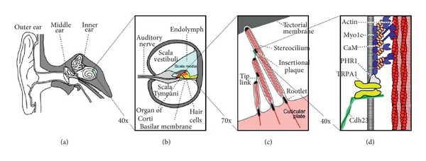
Figure 4.

Illustrations of the structure of the inner ear at various levels of magnification. The position of the inner ear in the temporal bone is shown in (a). The cross-sectional structure within one turn of the cochlea is shown in (b) with the fluid chambers separated by the basilar membrane and the organ of Corti. The details of the bundle of stereocilia that protrude from the top of the hair cells within the organ of Corti are shown in (c). Finally (d) shows the molecular details of the myosin motors that maintain the tension in the tip links that connect the individual stereocilia within the bundle. The transduction channels (here labelled TRPA1) are now believed to reside at the bottom end of the tip link rather than the top [47] (reprinted from Neuron, 48, LeMasurier and Gillespie, Hair-Cell Mechanotransduction and Cochlear Amplification, 403–415, Copyright (2005), with permission from Elsevier).
