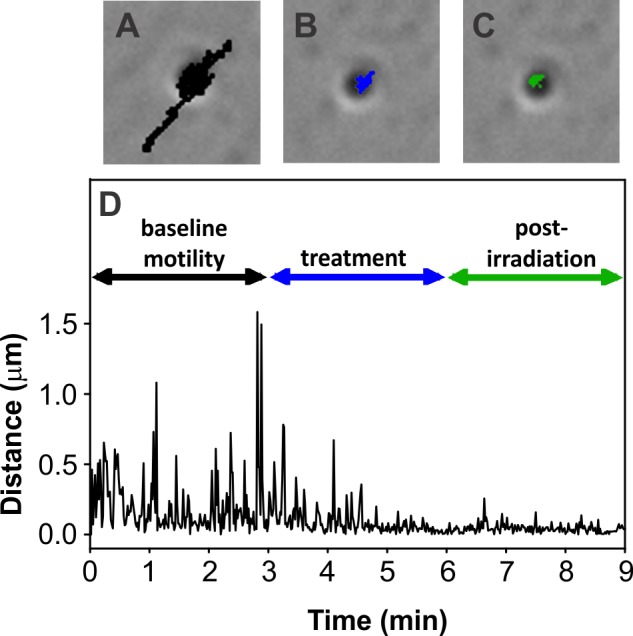
Figure 2.

Illustration of the movement of a representative lipofuscin granule at baseline and with sublethal blue light treatment. Bright field micrographs, with overlaid movement tracks, show organelle motility over 3-minute intervals at baseline ([A], track shown in black), then during ([B], track shown in blue) and after ([C], track shown in green) blue light irradiation. (D) Graphical representation of the movement of the lipofuscin granule shown in (A–C). Age of RPE donor: 84 years. Magnification for (A–C): ×600.
