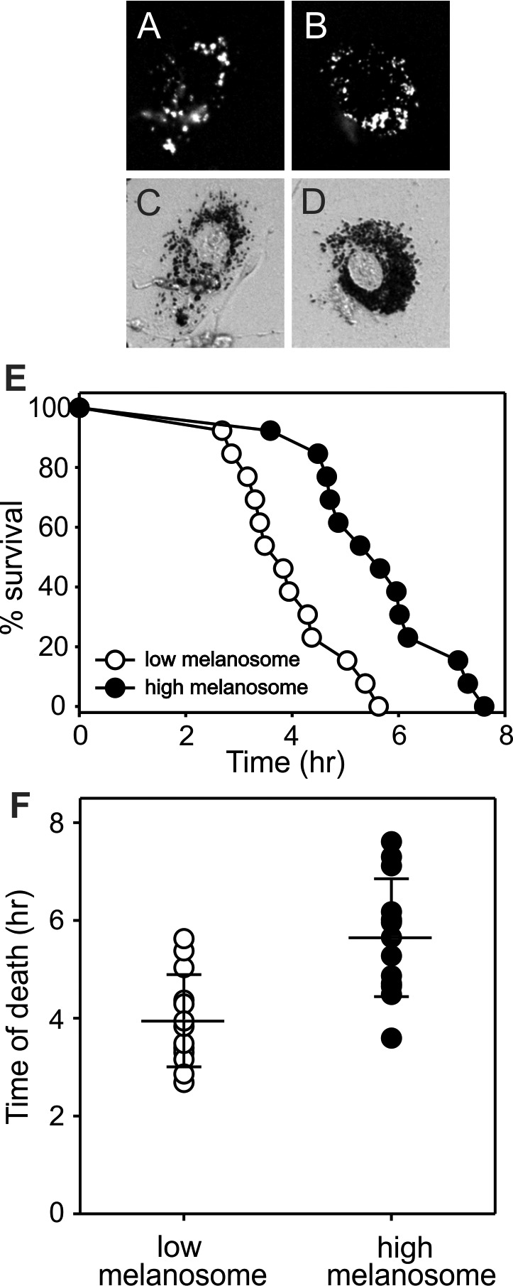
Figure 5.

Human RPE cells containing endogenous lipofuscin granules and differing numbers of phagocytized porcine melanosomes. Paired fluorescence (A, B) and bright field micrographs (C, D, respectively) illustrate cells with comparable lipofuscin and either low (C) or high (D) melanosome content. (E) Survival curves for RPE cells with low and high melanosome content during irradiation with blue light. Each dot represents an individual cell. (F) Demonstration of the mean time of cell death for cells with low and high melanosome content. Cell death was significantly later than for cells with higher melanosome numbers (P < 0.05, t-test). Wide cross bars indicate means, shorter error bars indicate SD. Age of RPE donor: 37 years.
