
Figure 1.
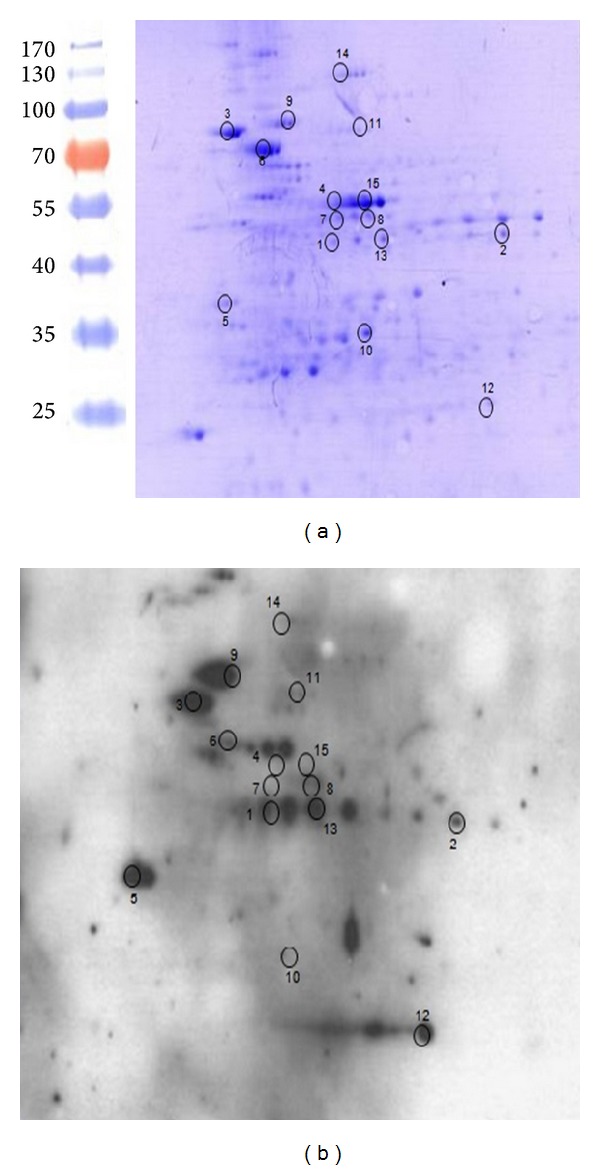
Analysis of MAP-cell wall proteins by 2D-SDS-PAGE. The cell wall protein fraction (CW) of MAP was resolved by 2D-SDS PAGE per duplicate and the resulting gels were (a) stained with Coomassie blue or (b) transferred onto a nitrocellulose membrane and subjected to Western blot. Sera from 5 positive animals were pooled and diluted to 1 : 100 to detect immunogenic proteins from the CW fraction. Molecular weight standards are shown on the left.