Figure 6.
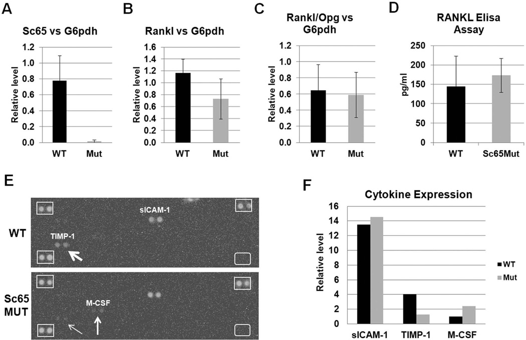
Measurements of RANKL, Opg and other cytokines. qPCR analysis showed reduction of Sc65 (A) but no significant changes in either RANKL (B) or osteoprotegerin (Opg) transcripts or their ratio (C) in cDNA from Sc65Mut or WT primary osteoblasts. Similar results were obtained when normalized against multiple housekeeping genes, including Gapdh, b-actin and G6pdh. D. Elisa assay for serum RANKL did not reveal significant differences between WT and Sc65Mut adult (4–6 month-old) male mice (n = 7–8, p = 0.3). E. Image of the Cytokine Array panels hybridized with pooled sera from Sc65Mut or WT mice (n=3) showing differential expression of both TIMP-1 and M-CSF between the two genotypes. Differences of expression are quantified by densitometry and shown in F.

