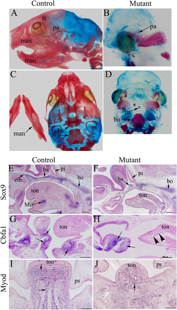
Fig. 2.
NCC-disruption of Dicer leads to abnormal skeletogenesis and muscle development in the craniofacial region. (A–D) Control (A, C) and mutant (B, D) head skeletons from newborn mice were stained with Alizarin red and Alcian blue. Head skeletons in the mutant mice were either missing or rudimentary. (E–H) In situ hybridization analysis was performed on sagittal sections of control (E, G) and mutant (F, H) embryos at E11.5 using probes against Sox9 (E, F) and Cbfa1 (G, H). Meckel’s cartilage and anterior basicranium (labeled by Sox9) were absent in mutants (E, F). Osteoblasts, which express Cbfa1, were absent in the mandible of mutants (indicated by arrow heads) (G, H). Arrows indicate positively stained cells. (I, J) In situ hybridization analysis was performed on transverse sections of control (I) and mutant (J) embryos at E13.5 using a probe against MyoD. In contrast to the striped pattern of muscle precursors observed in the primitive tongue of control animals, no obvious organization of muscle precursors was detected in mutant embryos. Arrows indicate examples of positively stained cells. bo, basioccipital; bs, basiosphenoid bone; eth, ethmoid bone; fr, frontal bone; man, mandible; max, maxilla; Mc, Meckel’s cartilage; pa, parietal bone; pi, pituitary gland; ps, palatal shelf; ton, tongue; Scale bar: 500 µm.

