
Figure 3. Effect of mutations on the binding of CovR and CovR-P to Pska.
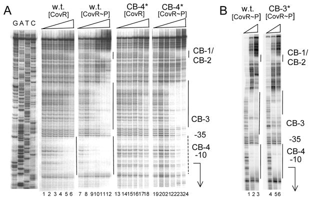
A: mutation in CB-4. Lanes 1–6 and 13–18: wild type DNA; lanes 7–12 and 19–24: mutant DNA with a TT to GG transversion in site CB-4 (denoted CB-4*). Lanes 1–6 and lanes 7–12: unphosphorylated CovR at 0, 0.25, 0.5, 1, 2 and 3 μM respectively. Lanes 13–18 and lanes 19–24: phosphorylated CovR at 0, 0.25, 0.5, 1, 2 and 3 μM respectively. The locations of the CovR binding sites CB-1 throgh CB-4 are indicated at the right of the figure. The solid lines indicate regions of DNaseI protection and the dashed line indicates partial DNaseI protection. The positions of the −10 and −35 regions of the promoter are indicated, and the arrow indicates the start point and direction of transcription. B: mutation in CB-3. Lanes 1–3: wild type DNA. Lanes 4–6: Mutant DNA with a TT to GG transversion in site CB-3 (denoted CB-3*). Lanes 1–3 and lanes 4–6; phosphorylated CovR at 0, 3, and 6 μM respectively. The location of the CovR binding sites CB1–CB4 are indicated at the right of the figure. The solid lines indicate regions of DNaseI protection. The positions of the −10 and −35 regions of the promoter are indicated, and the arrow indicates the start point and direction of transcription.