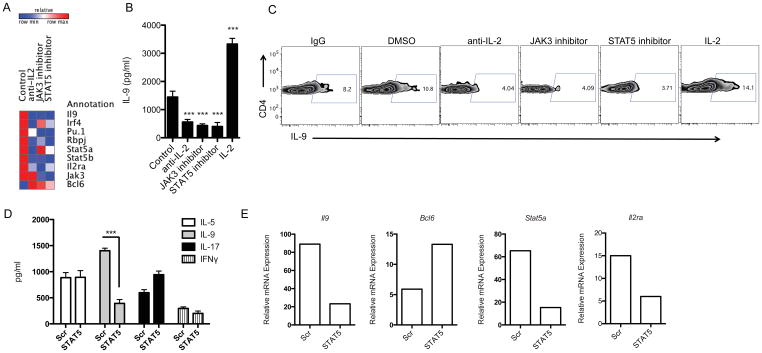
Figure 3. Opposite Role of STAT5A and BCL6 in the Regulation of Th9 Cell Development.
(A, B, C) Inhibition of IL-2/JAK3/STAT5 signaling decreases IL-9 production in Th9 cells. (A) Heatmap of gene regulated in Th9 cells treated with anti-IL-2 neutralizing antibody, and JAK3 and STAT5 inhibitors. Naive CD4+ T cells from WT mice were differentiated into Th9 cells in the presence or absence of the indicated treatment and gene expression was measured by Taqman PCR. (B, C) IL-9 protein expression by bead-based Luminex assay (B) as well as by intracellular flow cytometry (C) in treated Th9 cells is shown. (D, E) STAT5 knockdown reduces IL-9 and up-regulated BCL6 expression in Th9 cells. Naïve CD4+ T cells transfected using Amaxa with siRNA specific for STAT5A or scrambled (Scr) siRNA followed for polarization under Th9 cell conditions for 4 days. (D) Cytokine analysis of IL-5, IL-9, IL-17 and IFNγ expression in the supernatants was assessed by Luminex. (E) A representative gene expression of Il9, Bcl6, Stat5a and Il2ra in Th9 cells that were transfected with siRNA specific for STAT5A, or scrambled siRNA and measured by Taqman PCR. Data are representative of three experiments with similar results. ***p < 0.0001.

