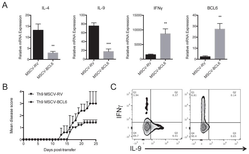
Figure 5. BCL6 Regulates Th9 Cell Pathogenicity in EAE mice.
(A) A representative gene expression of IL-4, IL-9, IL-17A, IFNγ and BCL6 in Th9 overexpressing BCL6 and control cells. MOG35–55-specific CD4+ T cells were isolated from 2D2 mice and were transduced with MSCV-BCL6-GFP retrovirus virus (RV) or with control empty vector (MSCV-RV) followed by in vitro polarization under Th9 conditions for 4 days in the presence of MOG35–55 peptide (20 μg/ml) and irradiated antigen-presenting cells. Cells were lyzed and analyzed for gene expression by Taqman. (B) A representative EAE experiment showing clinical scores (means ± s.e.m.) in mice after adoptive transfer of MOG35–55-stimulated 2D2 polarized under Th9 cell conditions with or without BCL6 overexpression (10 mice/group). Rag2−/− mice received MSCV-BCL6-transduced Th9 cells or control cells followed by immunization with MOG35–55 peptide (100 μg/ml). (C) Intracellular staining of transferred 2D2 cells. Infiltrated cells were prepared from the spinal cords of recipient mice (3 mice/group) followed by flow cytometry intracellular staining of IFNγ and IL-9. Data are representative of two experiments with similar results. **p < 0.01; ***p < 0.001.

