
FIG. 3.
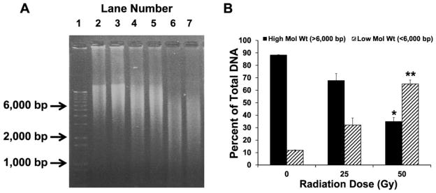
Effect of increasing doses of gamma radiation on calf thymus DNA fragmentation. DNA exposed to gamma radiation generates fragments of small molecular weights, which move faster than the high-molecular-weight DNA. Determining the density of the low-molecular-weight DNA fragments (<6,000 bps) compared to the high-molecular-weight DNA (>6,000 bps) reflects the extent of radiation-induced damage. Panel A: Lane 1: 1 kb DNA standard ladder; lanes 2 and 3: untreated calf thymus DNA; lanes 4 and 5: DNA exposed to 25 Gy; and lanes 6 and 7: DNA exposed to 50 Gy of radiation. Panel B: High- and low-molecular-weight DNA forms are shown as percentage of total DNA. For each condition, all samples were run in duplicate. The data are shown as mean ± standard deviation. P < 0.05 was considered significant. *,**Indicate significant differences compared to the untreated forms, respectively.