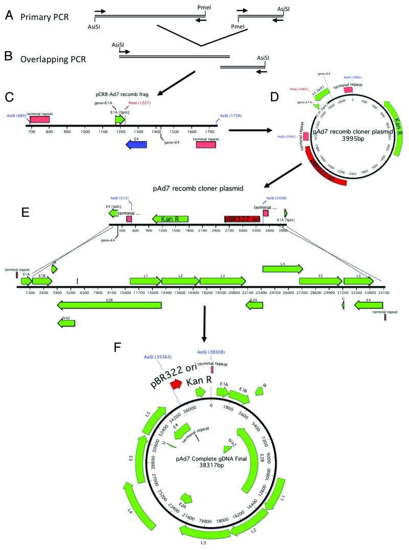
Figure 7. Schematic of Ad7 gDNA cloning. The right and left arms of Ad7 were amplified with primers designed to incorporate a unique PmeI restriction site, an overlap of ~27 nucleotides and flanking AsiSI restriction sites (A). The 2 PCR products were fused together in a second round of amplification (B). A schematic representation of the cloning fragment is shown (C). The cloning PCR fragment is ligated to the Kanamycin resistance gene and the pBR322 origin of replication (D). The recomb cloner plasmid was digested and recombined with the Ad7 genomic DNA in vitro using BJ5183 electrocompetent cells (E). The complete Ad7 genomic DNA plasmid is shown (F).

An official website of the United States government
Here's how you know
Official websites use .gov
A
.gov website belongs to an official
government organization in the United States.
Secure .gov websites use HTTPS
A lock (
) or https:// means you've safely
connected to the .gov website. Share sensitive
information only on official, secure websites.
