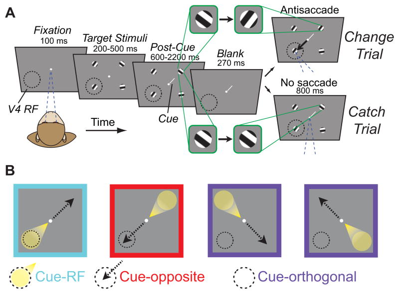
Figure 1. Cued change-detection and antisaccade task.
A, Task design and trial sequence. Monkeys fixated a white dot while four peripheral oriented-grating stimuli were presented. After a variable delay, stimuli disappeared then reappeared, either with or without one of the four stimuli rotating (change trial or catch trial, respectively). Monkeys could earn a reward by making a saccade to the diametrically opposite stimulus from the change on change trials, or by maintaining fixation on catch trials. A small, central cue (white line) indicated which stimulus, if any, was most likely to change. Green outlined panels emphasize the change in orientation, or lack of change, across the blank period. Dashed circle indicates area V4 receptive field (RF) locations and arrow indicates saccade direction; these were not visible to the monkey. All graphical elements are not precisely to scale; in particular, the cue is shown much larger than scale for visibility. B, Task conditions. On cue-RF trials, the relevant visual stimulus was in the RF of recorded neurons (spotlight) while the direction of the potential antisaccade was to the diametrically opposite stimulus (dashed arrow). Conversely, on cue-opposite trials, antisaccades were directed to the RF stimulus, while the relevant stimulus was diametrically opposite. On cue-orthogonal trials, neither the relevant stimulus nor the saccade target was in the RF.

