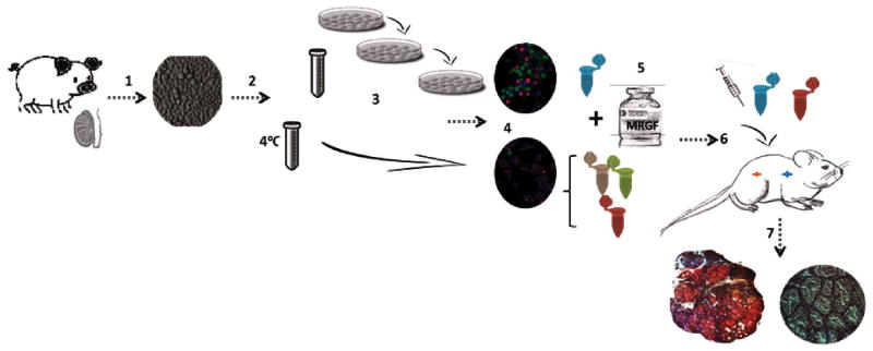
Figure 1.
Experimental design. 1 One week old piglet testes were enzymatically digested into a single cell suspension. 2 Cell suspension was split into two fractions: one was kept at 4°C until characterization and the other was used for enrichment. 3 Enrichment for germ cells by 3 sequential platings. 4 Both cell fractions were characterized by immunocytochemistry. 5 Cell pellets with the defined concentrations of germ cells were created and Matrigel was added (groups 2, 3 and 4). 6 Surgical grafting was performed. 7 After 24 and 40 weeks, the de novo formed tissue was harvested from host mice and cross sections were analyzed for the quantification of de novo formed tubules, detection of UCHL-1+ cells and development of spermatogenesis.

