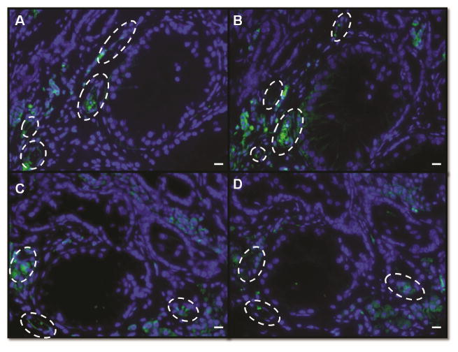
Figure 2.
Validation of alpha smooth muscle actin as a marker to identify blood vessels. Immunofluorescence from sequential cross sections from grafts recovered after 12 weeks labeled against alpha smooth muscle actin and CD31, punctuated white lines indicates blood vessels. A, C Grafts stained against alpha smooth muscle actin. B, D Grafts stained against CD31. Bar=50μm.

