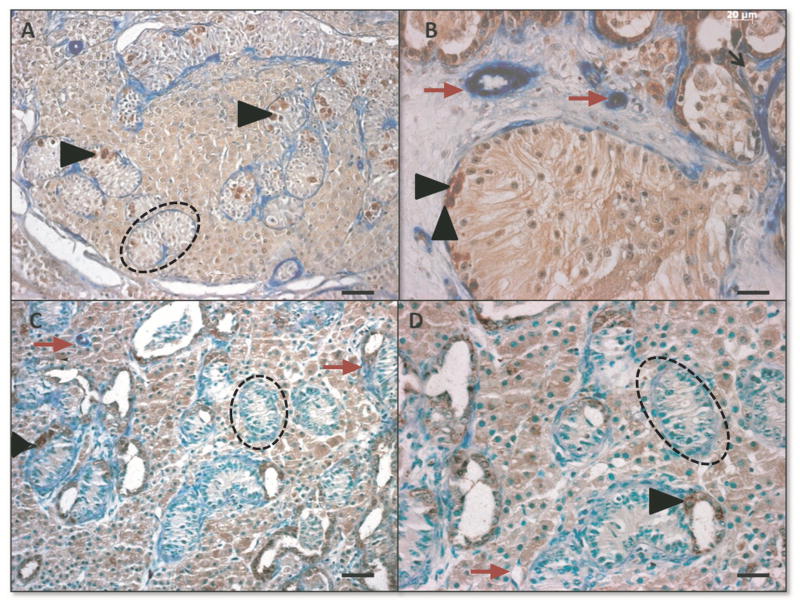
Figure 3.
Morphology and immunohistochemistry to identify UCHL1+ (brown, black arrow heads) and alpha-smooth muscle actin + (blue, red arrows) cells in de novo formed testis tissue from VEGF experiment, punctuated black lines identify seminiferous tubules. A VEGF-165 treated tissue; bar=50μm, B: VEGF-165 treated tissue; bar=20μm, C Control tissue; bar=50μm; D Control tissue; bar=20μm.

