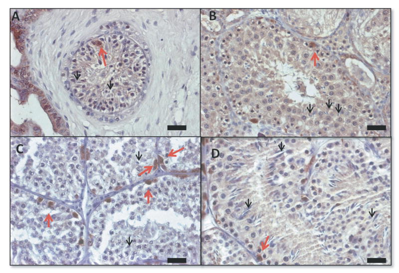
Figure 5.
Morphology of de novo formed tubules and spermatogenesis by immunohistochemistry for the detection of UCHL1 40 weeks after transplantation. A Control, group 1: 50×106 cells, B group 2: 50×106 cells in Matrigel, C group 3:10×106 enriched with spermatogonia in Matrigel, D group 4:10×106 cells in Matrigel. Red arrows indicate undifferentiated spermatogonia and black arrows point to elongated spermatids. Bars=20μm.

