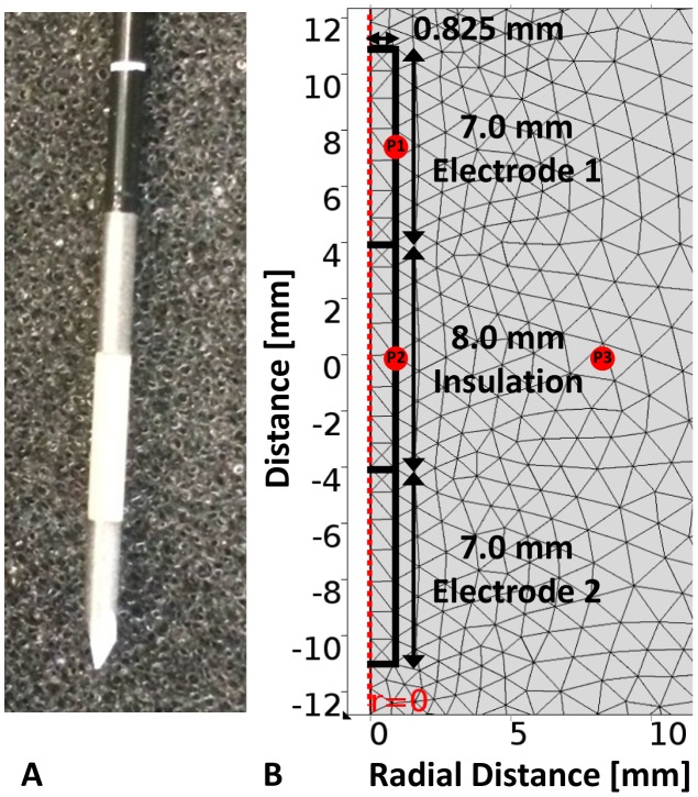
Figure 1. Physical electrode and mesh visualization for the extremely fine setting used in the numerical modeling software.
Photograph of the physical bipolar electrode (left) and the electrode domain (right) geometry with the corresponding mesh employed in the computational modeling of the bipolar probe used in irreversible electroporation of liver tissue. The points P1, P2, and P3 depict the arbitrary locations at which the temperature was evaluated to determine the length required for thermal equilibration post-treatment.

