
Figure 3.
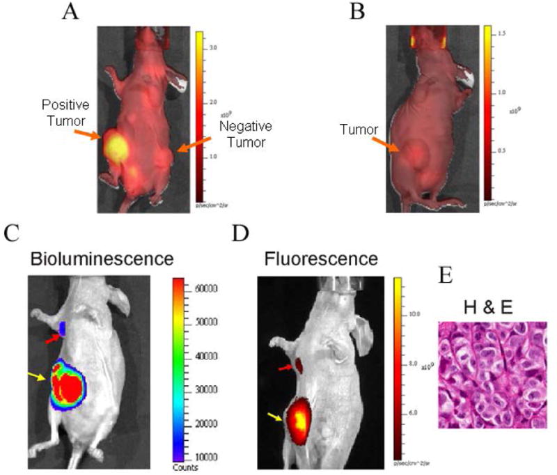
In vivo selectivity and representative in vivo images of spontaneous metastasis into the axillary lymph node by luciferase expressing ZR-75.1 human breast cancer cells. (A) In vivo fluorescence image of mouse bearing positive (ZR-75.1) and negative (MDA-mb-231) xenograft tumors, 24 h post injection of agent. Autofluorescence background signal is observed throughout the animal. Note the bright signal in the positive tumor and that agent has cleared from the entire mouse, including the negative tumor at this time-point. (B) Fluorescence image of a ZR-75.1 MFP tumor bearing mouse that was preinjected with 250 μg unlabeled mammaglobin-A monoclonal antibody, and 4 h later followed by injection of 50 μg MamAb-680. The image was acquired 24 h after injection with labeled probe. (C) Bioluminescence image of luciferase activity in the primary tumor (yellow arrow) and axillary lymph node metastasis (red arrow). (D) After peritumoral injection (24 h) of MamAb-680, the same mouse was imaged for fluorescence, and signal was observed to emit from the mammaglobin-A expressing (ZR-75.1) axillary lymph node metastasis (red arrow) and primary tumor (yellow arrow), following clearance of MamAb-680 from the MFP. (E) H & E staining of the ALN metastasis from the same mouse. It was noted that the shape of the metastasis matched the elongated shape observed in both the bioluminescence and fluorescence images.