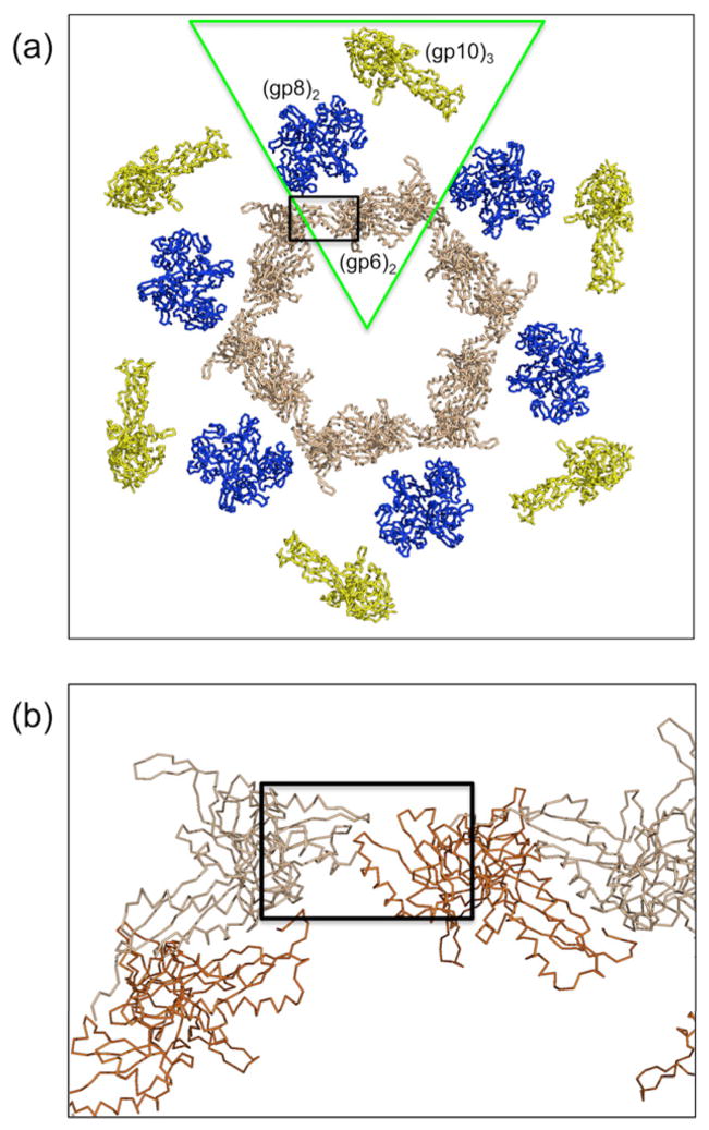
Fig. 5. Association of the proteins in the in vitro assembled baseplate.
(a) The hexameric ring of the gp10 (yellow), gp8 (blue) and gp6 (beige) proteins, based on the earlier cryoEM reconstruction of the star shape baseplate and the crystal structures of trimeric gp10, dimeric gp8, and dimeric gp6, around the 6-fold crystallographic axis. A wedge corresponds roughly to the green triangle. (b) There is a clash (black rectangle) between neighboring ends (brown and beige) of the 6-fold related gp6 molecules when fitting the independently determined crystal structures into the cryoEM density, indicating that there are at least some small conformational differences in the crystal structures.

