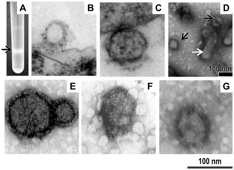
Figure 2. Photograph of a sucrose gradient and transmission electron microscopy (TEM) of virus-like particles (VLPs).
The culture supernatant of Sf21 insect cells at 72-h post-infection with recombinant baculoviruses (rBVs) was subjected to sucrose gradient centrifugation; the opalescent band (A, indicated by the arrow) was collected for TEM after staining with 1% uranyl acetate. a trace of residual baculoviruses(D, indicated by the white arrow) were consistently detected in VLP preparations(D, indicated by the black arrow). Both PPRV-H VLPs (F) and PPRV-F VLPs (G) isolated in this manner showed spherical shapes upon TEM, with spikes on their surfaces, and with a diameter of approximately 80–100 nm were observed. Native peste des petits ruminants virus (PPRV) propagated in Vero cells was also purified and negatively stained (E). Ultrathin sections of Sf21 insect cells showing budding of PPRV-H and PPRV-F VLPs (B and C) from the plasma membrane at 48 h after infection with rBVs.

