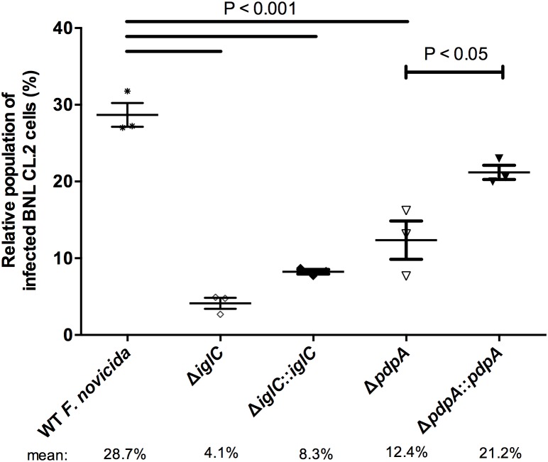
Figure 3. F. novicida lacking either IglC or PdpA reduces liver epithelial cell colonization.
Samples were fixed at 24 h PI, differentially stained for intracellular and extracellular bacteria, and then visualized by fluorescence microscopy. The proportion of infected cells was tallied from over 1,000 cells. Cells containing one or more intracellular bacteria are considered ‘infected’. Error bars, S.E.M. (n = 3).

