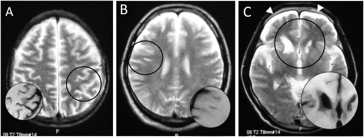
Figure 1. Magnetic resonance images of brains of three Ama divers: hyperintense area on T2-weighted image (circle), corresponding to hypointensity on T1-weighted image (inset).
A patchy shadow in the left parietal cortex (A, No. 2), a linear subcortical lesion in the right frontal lobe (B, No. 5), and deformity of bilateral caudate heads and subdural fluid collection (allow heads) (C, No. 11).

