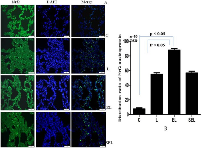
Figure 4. Immunofluorescence assays of nuclear localization of Nrf2 protein using fluorescence microscope (original magnification×400).
“A” showed the pictures of immunofluorescence staining, while “B” presented the distribution ratios of Nrf2 nucleoprotein to the number of nuclei in unit area of five fields among four groups. Green standed for Nrf2-FITC stained sections, while blue standed for images of DAPI stained nuclei. It was confirmed that Nrf2 increasingly translocated from cytoplasm into the nucleus by electroacupuncture protocols (P<0.05) rather than sham electroacupuncture stimulation (P>0.05). Data were representative of three independent experiments. Values were mean ± SD, and ten control, LPS, EL and SEL experiments were performed for each group.

