Figure 2.
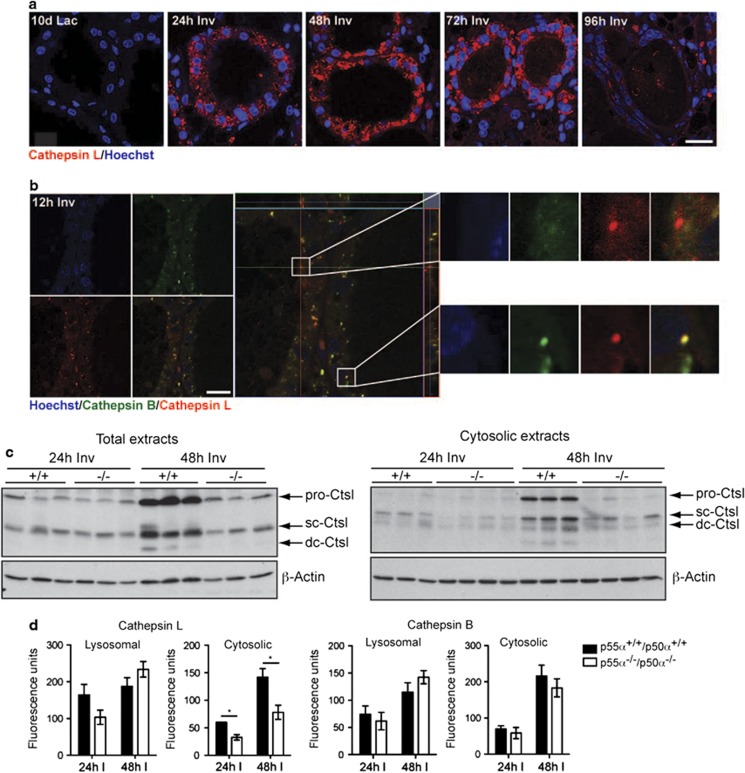
p55α/p50α regulate cathepsin L expression and cytosolic activity. (a) Rapid upregulation of cathepsin L at the onset of involution. Immunohistochemical analysis of cathepsin L (red) in control mice. 10d Lac, 10 days lactation; 24, 48, 72, 96 h Inv, hours of involution. Nuclei are stained with Hoechst. Scale bar, 20 μm. (b) Vesicles containing cathepsin L (red) but not cathepsin B (green) are shown by immunohistochemistry on 12-h involution control samples. Insets of an example of a vesicle containing only cathepsin L and one containing both cathepsin L and B are shown on the right side. Nuclei are stained with Hoechst. Scale bar, 20 μm. (c) Left, immunoblot showing decreased levels of cathepsin L (Ctsl) in the p55α−/−/p50α−/− (−/−) mice as compared with control p55α+/+/p50α+/+(+/+) mice. Three individual mice are shown per genotype at both 24-h involution and 48-h involution. Right, immunoblot of cytosolic extracts of three control and four p55α−/−/p50α−/− mammary glands (sc, single chain; dc, heavy chain of the double-chain form; both the single-chain and the double-chain form are active). β-Actin is shown as loading control. (d) Reduced cytosolic activity of cathepsin L at 24- and 48-h involution in p55α−/−/p50α−/− glands when compared with controls, as opposed to unchanged cathepsin B activity shown by subcellular fractionation and subsequent cathepsin activity measurement. All values are means±S.E.M. of at least three independent biological repeats. (*P<0.05, as determined by Student's t-test)

