Figure 3.
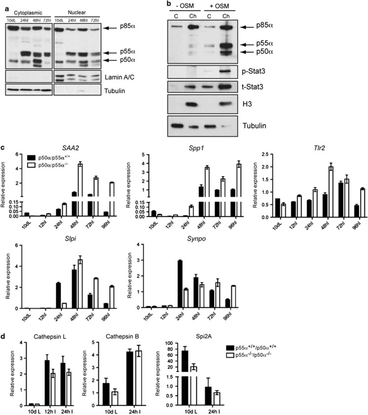
p50α and p55α localize to the nuclei of mammary glands during involution and regulate transcription of a subset of genes. (a) Nuclear/cytosolic fractionation of mammary glands showing both cytosolic and nuclear localization of p50α, p55α and p85α. Lamin A/C and tubulin are shown as loading and purity controls for the nuclear and cytosolic fraction, respectively. (b) Chromatin-enriched extracts of EpH4 mammary epithelial cells treated with vehicle (−OSM) or OSM (+OSM) showing the presence of phospho-Stat3 (p-Stat3) and p55α/p50α mainly in the chromatin-enriched fraction (ch) of the OSM-treated cells as opposed to the cytosolic (C) fraction, whereas p85α is present on chromatin also in the control cells. t-Stat3, total Stat3. Histone H3 (H3) is shown as chromatin marker, whereas tubulin shows the cytosolic fraction. Notably, tubulin contamination in the chromatin fraction of the OSM-treated cells is almost undetectable. (c) Quantitative real-time PCR relative to cyclophilin a of the following genes: SAA2, serum amyloid A 2; Spp1, secreted phosphoprotein 1; Tlr2, Toll-like receptor 2; Slpi, secretory leukocyte protease inhibitor; Synpo, synaptopodin. All results are means±S.E.M. for three independent measurements and are representative of two independent biological repeats. (d) Quantitative real-time PCR relative to cyclophilin a of Cathepsin L and B and Spi2A expression at the indicated time points of mammary gland involution showing a decreased, although not significant, expression of cathepsin L in the p55α−/−/p50α−/− glands as compared with the controls. Results are means±S.E.M. for at least four independent biological repeats

