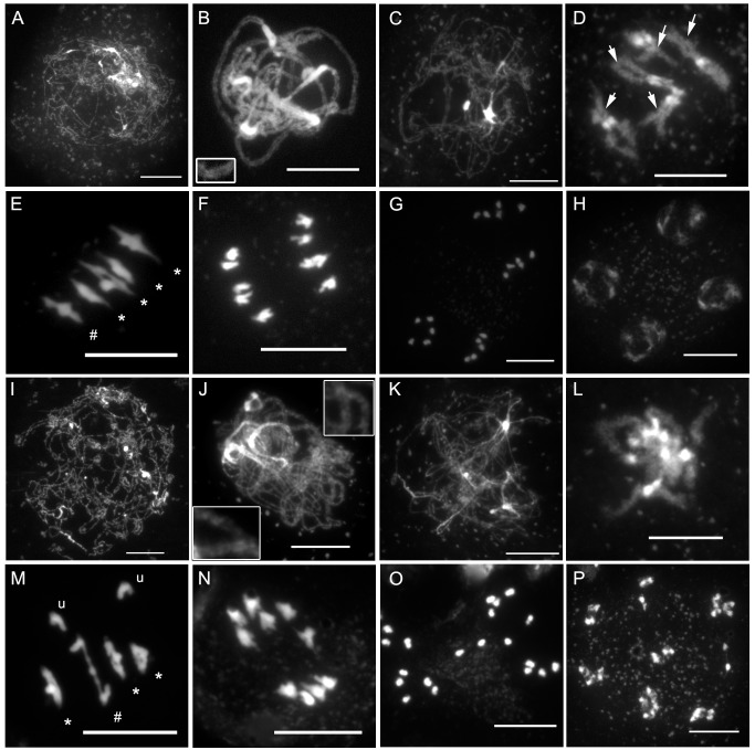
Figure 3. axr1 mutants show normal meiotic progression but reduced bivalent formation at metaphase I.
DAPI staining of meiotic chromosomes in wild type (A–H) and axr1 (N877898, I–P). At the onset of meiotic prophase I (A and I), chromosomes can be identified. Chromosome alignment and synapsis then proceeds, leading eventually to the pachytene stage in wild type (B), where homologous chromosomes are synapsed along their entire length. This association can be observed in axr1 (J, enlarged regions) but remains partial. Then, the SC disappears at diplotene (C and K), condensation proceeds, and bivalents can be identified in wild type at diakinesis (D), but this stage is rarely observed in axr1 (L). At metaphase I, the five Arabidopsis bivalents can be identified in wild type (E), segregating at anaphase I (F). In axr1, a mixture of bivalents and univalents are observed (M), leading to subsequent improper segregation at anaphase I (N). Sister chromatids segregate at meiosis II (G and O), leading to balanced tetrads in wild type (H), unbalanced tetrads (not shown) or polyads in axr1 (P). At metaphase I, univalents (u) can be distinguished from ring bivalents (where a chiasma occurred in each of the two chromosome arms, *) and from rod bivalents (where only one chromosome arm shows a chiasma, #). Arrows in (D) indicate some of the chiasma-containing arms. Bar, 10 µm.

