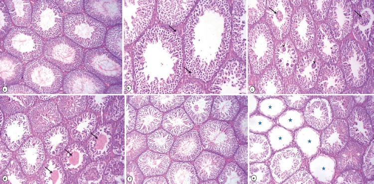
Figure 1.

Photomicrograph of rat testes stained with HE: Rats received lipoic acid (1%) showing normal histoarchitecture of testes (a X.160). Rats received acrylamide (0.05%) exhibited vacuolar degeneration of the germinal epithelium and Sertoli cells (arrows) (b X.250), sloughing of the germinal epithelium (arrows) with giant cell formations (arrowheads) in the lumen of seminiferous tubules (c X.160), depletion of germinal cells and hyalinization of the luminal contents (arrows) (d X.160) and some seminiferous tubules loss of all germinal epithelium cells (stars) (e X.160). Improvement of spermatogenesis in testes of rats treated with acrylamide + lipoic acid (f X.160).
