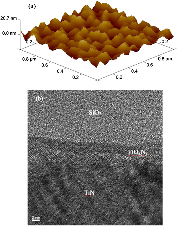
Figure 3.

AFM and HRTEM images for TiN layer. (a) Atomic force microscope (AFM) image shows surface roughness of TiN layer with a scan area of 1× 1 μm2. (b)The TiN surface is oxidized and is observed by high-resolution transmission electron microscope (HRTEM) image.
