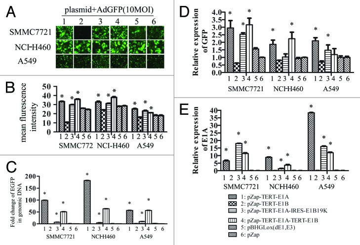
Figure 5. Differential effect of different element of E1 genes on AdGFP replication. SMMC7721, NCI-H460, and A549 cells grown on 6-well plates were transfected with 2 µg of plasmid DNAs using lipofectamine 2000, empty vector pZap was used as control. Twenty-four hours after transfection, cells were infected with 10 MOI of AdGFP. Four hours later AdGFP were removed and cells were washed 3 times using PBS and cultured with normal medium. The representative pictures of GFP expression were taken by epi-fluorescence microscopy 24 h later after infection (A). A summary of FACS analysis for GFP expression in cells mentioned above (B). GFP DNA detected by quantitative PCR (C). GFP mRNA detected by quantitative RT-PCR (D). E1a mRNA detected by quantitative RT-PCR (E).

An official website of the United States government
Here's how you know
Official websites use .gov
A
.gov website belongs to an official
government organization in the United States.
Secure .gov websites use HTTPS
A lock (
) or https:// means you've safely
connected to the .gov website. Share sensitive
information only on official, secure websites.
