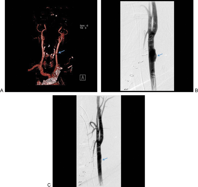
Figure 3.

(A) A grade I lesion identified as a luminal narrowing of the left common carotid is seen on the reconstructed CTA. (B) DSA demonstrates progressive worsening of the common carotid injury. (C) Endovascular intervention with a stent was placed successfully. The arrows indicate the site of grade I carotid injury before and after endovascular stenting. CTA, computed tomography angiography; DSA, digital subtraction angiography.
