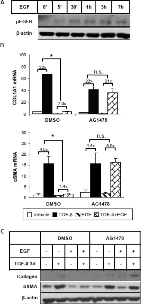
Figure 2.
EGF represses TGF-β-induced collagen and αSMA expression through EGFR. (A) HKCs were treated with EGF (25 ng/ml) for various times as indicated. Phospho-EGFR was examined by western blot. β-actin was used as a loading control. (B) Cells were pretreated with DMSO or EGFR tyrosine kinase inhibitor AG1478 (5 μM) for 1 hour, followed by incubation with TGF-β (1 ng/ml) and/or EGF (25 ng/ml) for 24 hours. COL1A1 and αSMA mRNA levels were examined by quantitative PCR. *p<0.05 fold induction by TGF-β comparing with and without EGF treatment (n=3). (C) Cells were treated as above but for 72 hours. COL1A1 and αSMA protein levels were examined by western blot. β-actin was used as a loading control.

