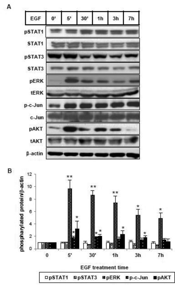
Figure 3.
EGF activates multiple signaling pathways in HKC. (A) Cells were incubated with EGF (25 ng/ml) for the indicated intervals. Total cell lysates were analyzed by western blot using specific antibodies against phosphorylated forms of STAT1, STAT3, ERK, c-Jun, AKT (pSTAT1, pSTAT3, pERK, p-c-Jun, pAKT) and their corresponding non-phosphorylated forms. β-actin was used as a loading control. (B) Densitometric analysis of pSTAT1, pSTAT3, pERK, p-c-Jun and pAKT was normalized to β-actin and expressed as fold change over vehicle alone. *p<0.05, **p<0.005 comparing with vehicle control (n=3).

