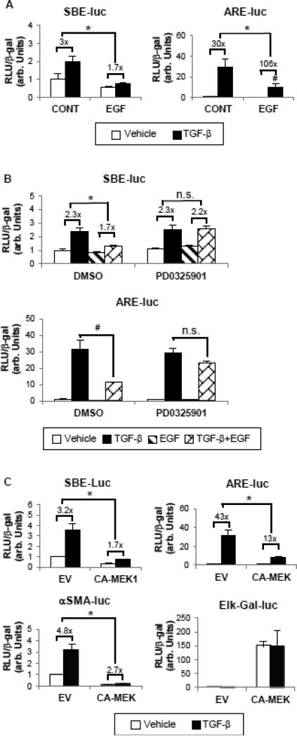
Figure 5.
EGF decreases Smad2 and Smad3 transcriptional activities via ERK activation. (A) Cells were transfected with either ARE-Luc plus FAST-1 or SBELuc reporter constructs to detect the transcriptional activity of Smad2 and Smad3, respectively. 24 hours after transfection, cells were exposed to 1 ng/ml TGF-β with or without EGF (25 ng/ml) for another 24 hours. *p<0.05 fold induction by TGF-β comparing with and without EGF treatment. #p<0.05 comparing absolute activities induced by TGF-β in the presence or absence of EGF treatment (n=3). (B) Transfected cells were pretreated with DMSO or PD0325901 (100 nM) for 1 hour before incubation with TGF-β and/or EGF for 24 hours. *p<0.05 fold induction by TGF-β comparing with and without EGF treatment. #p<0.05 comparing absolute activities induced by TGFβ in the presence or absence of EGF treatment (n=3). (C) A constitutively active MEK1 construct (CA-MEK1, 0.5 μg) or its empty vector (EV) was overexpressed in HKCs along with ARE/SBE-Luc or αSMA promoter reporter construct (SMA-Luc). Elk-Gal-Luc was used to determine the ERK functional activity. *p<0.05 fold induction by TGF-β compared with EV group. In all experiments, the luciferase activity assayed in triplicates was normalized to β-galactosidase (β-gal) to control transfection efficiency.

