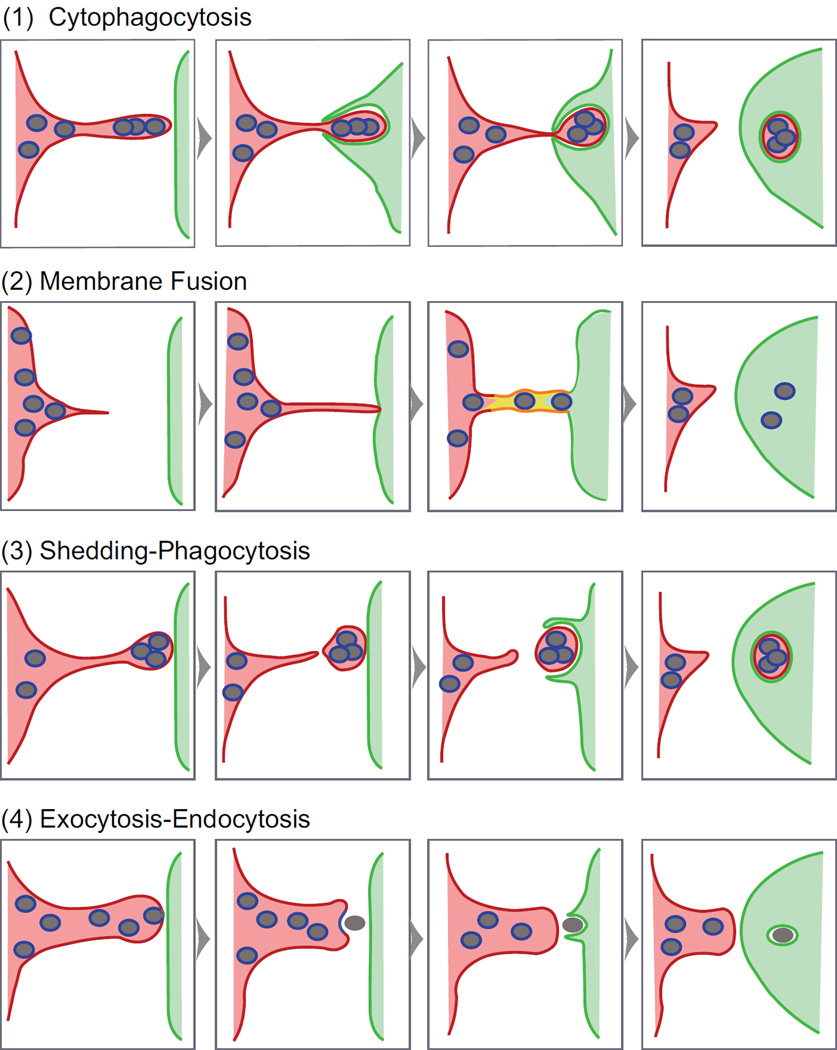
Figure 1. Melanosome transfer mechanisms.
Shown are four possible mechanisms of intercellular melanosome transfer. The cartoon, which is based off of a simpler cartoon in [7], uses color coding to mark the identity of the three membranes that are relevant to the characterization of transfer mechanisms: the limiting membrane of the melanosome (blue), the plasma membrane of the melanocyte (red) and the plasma membrane of the keratinocyte (green) (the yellow in Mechanism 2 indicates a mixture of these latter two membranes). Filopodia on the surface of the melanocyte could give rise to the intercellular bridge connecting the cells in Mechanism 2 [7, 10]. Importantly, three of the four mechanisms (Mechanisms 1, 3 and 4) employ phagocytosis by the keratinocyte to complete the transfer process. This step requires the expression on the surface of the keratinoctye of the protease activated receptor Par2 (see [27] for review). We note, however, that phagocytic extensions typically cannot “bite through” the object being engulfed [39], arguing against Mechanism 1 (see the text for other reservations regarding this mechanism).

