
Figure 3. Serial section and immuno-electron microscopy of human skin support the Exocytosis-Endocytosis mechanism.
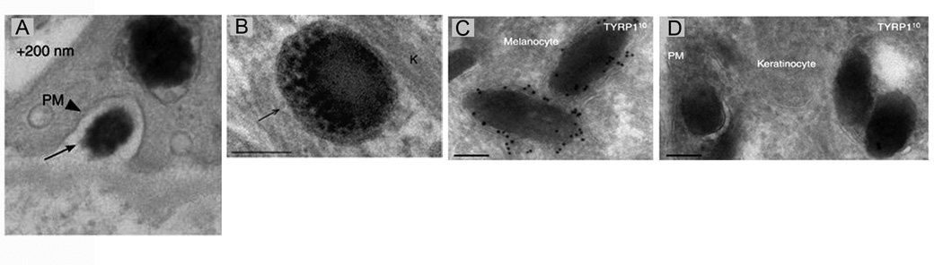
Panel A shows an image of a melanocore in the extracellular space between a melanocyte and a keratinocyte (this image is from a series of consecutive sections that together prove this melanocore is extracellular). Panel B shows the single membrane surrounding a melanocore inside a keratinocyte. Panel C shows the extensive immunogold labeling of a two melanosomes inside a melanocyte using an antibody to TYRP1, a glycoprotein present in the limiting membrane of the melanosome. Panel D shows the minimal immunogold labeling for TYRP1 on several melanocores inside a keratinocyte. Together, Panels C and D argue that the limiting membrane of the melanosome is “left behind” during transfer, consistent with Mechanism 4. Reproduced with permission from the Journal of Investigative Dermatology [24] ••.