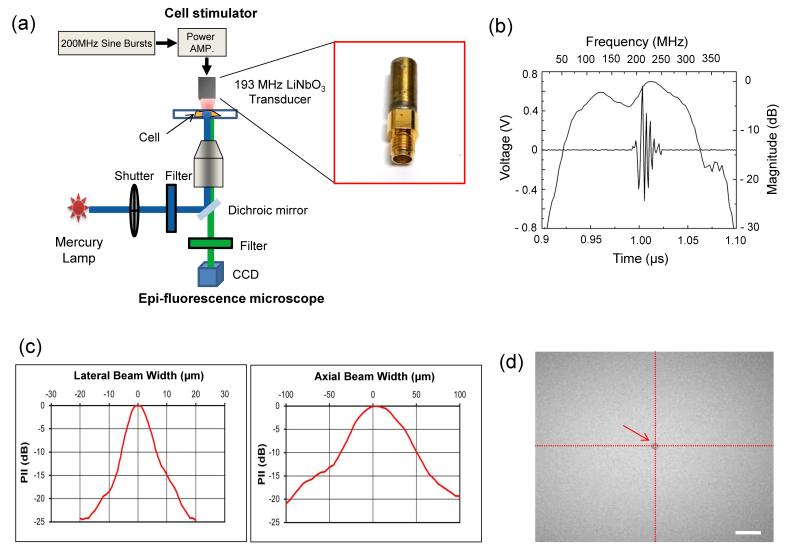
Fig. 1. Characterization of high-frequency ultrasound beams and the beam localization at the region of interest.
(a) A high-frequency ultrasound mechanotransduction system (b) Pulse-echo characteristics of the transducer (c) Lateral (left) and axial (right) beam profiles. PII stands for pulse intensity integral. (d) Localization of an ultrasound microbeam at the center of an image using acoustic tweezers. The arrow indicates a 5 μm microbead trapped at beam focus. The scale bar indicates 20 μm.

