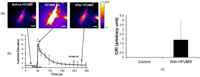
Fig. 2. Cytoplasmic Ca2+ changes in HUVECs due to HFUMS.
(a) Fluorescence images obtained before (left), during (middle), and after (right) HFUMS (input voltage: 15.8 Vpp, PRF: 1 kHz, and duty cycle: 1 %) (b) Ca2+ changes in HUVECs over times. HFUM was switched on at 60 s and off at 240 s (c) Quantitative analysis of calcium elevation. The CRI values for HUVECs without (control) (n=21) and with (n=21) HFUMS were calculated with the program described previously. The scale bar indicates 10 μm.

