
Figure 3.
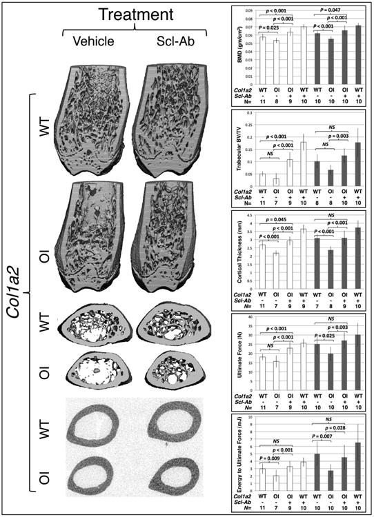
Sclerostin inhibiting antibody treatment improves bone properties in wild-type and in OI mice. (left panels) 3-D μCT reconstructions of the distal and midshaft femurs from male 12-week-old wild-type (Col1a2+/+) and OI (Col1a2+/p.G610C) mice that received 6 weeks of vehicle or Scl-Ab. (right panels) Graphs depicting mean (± SD) measures of bone mineral density (BMD), distal femur trabecular bone volume/total volume (BV/TV), midshaft femur cortical thickness, ultimate force to failure, and energy to ultimate force in female (open bars) and male (shaded bars) 12-week-old wild-type and OI mice that received 6 weeks of vehicle or Scl-Ab. Col1a2 genotypes, wild-type (WT) or heterozygous knockin (OI) are indicated, as is the number (N) of animals with each genotype that were studied. Brackets indicate comparisons and p values between vehicle treated OI and vehicle treated wild-type mice, between Scl-Ab treated OI and vehicle treated OI mice, and between Scl-Ab treated OI and vehicle treated wild-type mice. NS – not significant.