
Figure 1.
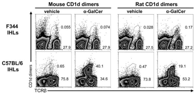
Identification of rat iNKT cells. IHLs derived from F344 inbred rats or C57BL/6 inbred mice were analyzed by flow cytometry using rat and mouse CD1d dimers loaded with α-GalCer or vehicle as control. Rat and mouse TCRβ expression was also analyzed. The secondary DαM Ab used to visualize the bound dimers also stained mouse IgG-positive B cells, thus these cells were excluded in the analysis of mouse IHLs by gating out CD19+ cells, which were 23% of all IHLs. Numbers indicate the percentages of the gated cells. The data shown are representative of three independent experiments.