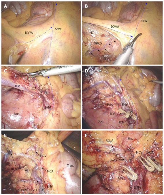
Figure 1.

D3 lymphadenectomy of laparoscopic extended right hemicolectomy. A: Locating the ileocolic and superior mesenteric vessel pedicles; B: Exploring the RRCS between the mesocolon and the pre-renal fascia; C: Dissecting the ileocolic vessels and SMV; D: Extending the RRCS with strict preservation of the mesopancreas and mesocolon; E: Dissecting the middle colic vessels, gastrocolic trunk, and its tributaries; F: Lymphadenectomy at the hypopyloric region. SMV: Superior mesenteric vein; ICV: Ileocolic vein; ICA: Ileocolic artery; MC: Mesocolon; RRCS: Right retrocolic space; RPRF: Right pre-renal fascia; Duo: Duodenum; Pan: Pancreas; GCT: Gastrocolic trunk; RGeV Gastroepiploic vein; RCV: Right colic vein; ASPDV: Anterior superior pancreaticoduodenal vein; MCV: Middle colic vein; MCA: Middle colic artery; Tran: Transverse colon; Sto: Stomach; RGeA: Gastroepiploic artery.
